Can Pregnancy Be Mistaken for a Fibroid on Ultrasound?

Summary
- The short answer: While rare, an early pregnancy can sometimes be confused with a degenerating fibroid on a very basic ultrasound, especially if the pregnancy is in its earliest days before a distinct sac forms.
- Visual differences: Fibroids are solid muscle tumours that appear dense and grey on a scan. A healthy pregnancy typically shows a dark, fluid-filled gestational sac containing a yolk sac or embryo.
- The role of hormones: A simple blood test measuring hCG (the pregnancy hormone) quickly clears up any confusion between a fibroid and a pregnancy.
- Expertise matters: High-resolution ultrasound equipment operated by an experienced gynaecologist eliminates the risk of misdiagnosis, ensuring you receive the correct care immediately.
If you are reading this, you might be feeling a little anxious, confused, or simply curious about an ultrasound result.
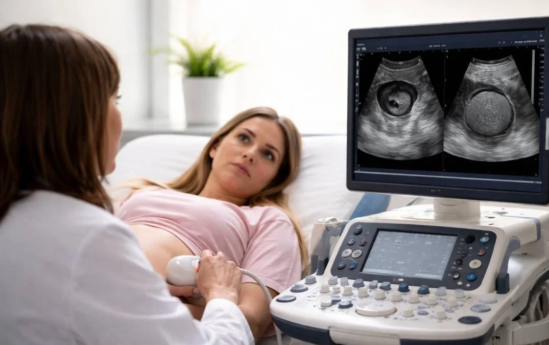
Sitting in the doctor’s office while looking at a grainy, black-and-white monitor can feel overwhelming.
When you see a shadow on the screen, your mind immediately races with questions.
One of the most surprising questions I hear in my clinic is whether a fibroid can be mistaken for a pregnancy, or vice versa.
When you are hoping to start a family or experiencing unexpected symptoms, you need absolute clarity.
Let us walk through exactly what happens during these scans, why confusion can occasionally happen, and how we ensure you get the accurate answers you deserve.
Why do we perform ultrasounds in early pregnancy?
Ultrasounds are our window into your reproductive health. We use sound waves to create images of your uterus, ovaries, and surrounding structures.
When you miss a period or have a positive pregnancy test, an early scan serves several vital purposes.
First, we need to confirm that the pregnancy is safely located inside the uterus.
We also check the heartbeat, estimate the gestational age, and ensure there are no underlying issues that could complicate your journey.
However, the uterus is a complex, muscular organ. It is not always an empty space waiting to be photographed.
Sometimes, the scan reveals unexpected structures, which brings us to the topic of fibroids.
What exactly is a uterine fibroid?
Before we compare the two, you need to understand what a fibroid actually is.
Uterine fibroids are non-cancerous growths made of smooth muscle cells and fibrous connective tissue.
They develop within the wall of the uterus. They are incredibly common, affecting a large percentage of women during their reproductive years.
Fibroids range in size from tiny seedlings that are undetectable to the human eye, to large masses that can alter the shape of your uterus.
Because they are solid masses of tissue, they interact with ultrasound waves differently than fluid or empty space.
Most women do not even know they have them until they show up on a routine scan.
Is it possible to mistake a pregnancy for a fibroid?
The direct answer is that it is highly unlikely for an experienced specialist using modern equipment, but it is technically possible under very specific circumstances.
Let me explain how this rare mix-up can occur.
If a woman has a very early pregnancy—perhaps just four or five weeks along—the gestational sac is minuscule.
At this stage, it might just look like a tiny thickening of the uterine lining.
On the other hand, if a woman has a small fibroid that is undergoing a process called “degeneration” (where it outgrows its blood supply and begins to break down), the centre of the fibroid can become cystic or fluid-filled.
To an untrained eye or on an older, low-resolution ultrasound machine, a small, fluid-filled degenerating fibroid might briefly mimic the appearance of a tiny gestational sac.
Conversely, a very early, poorly defined pregnancy might look like a small thickening or shadow, resembling a small fibroid.
However, this confusion is usually fleeting and easily resolved.
How do fibroids and pregnancy look different on a scan?
When I look at your ultrasound, I am evaluating the density, shape, and echoes of the sound waves.
Fibroids and pregnancies have very distinct acoustic signatures.
A fibroid is a solid tumour. Sound waves bounce off the dense muscle tissue, making it appear as a grey, solid, sometimes swirling mass on the screen.
It often casts a shadow behind it.
A pregnancy, however, begins with a gestational sac. Fluid does not reflect sound waves, so the fluid inside the sac appears completely black on the monitor.
Within that black space, as the weeks progress, we look for the bright white ring of a yolk sac, and eventually, the tiny fetal pole with a fluttering heartbeat.
A solid fibroid will never have a yolk sac or a heartbeat.
Could a misdiagnosis affect your health or baby?
Accuracy in gynaecological imaging is absolutely critical.
If a pregnancy is mistaken for a fibroid, you might not receive the necessary prenatal guidance right away.
You could also be subjected to unnecessary worry.
Alternatively, if a bleeding fibroid is mistakenly assumed to be a complicated pregnancy, you might face unnecessary medical interventions or anxiety about a miscarriage when the issue is entirely structural.
Furthermore, fibroids and pregnancy often coexist. Many women safely carry babies to term with fibroids present.
In these cases, we must accurately map the size and location of the fibroids to ensure they do not interfere with the baby’s growth or the delivery process.
How can we confirm what the ultrasound shows?
If there is ever a shadow of a doubt on the ultrasound screen, we never rely on guesswork. We have highly reliable methods to give you a definitive answer immediately.
The most powerful tool we have is the beta-hCG blood test. Human chorionic gonadotropin (hCG) is a hormone produced exclusively during pregnancy.
A fibroid will never produce hCG. If your ultrasound shows a confusing mass, a quick blood test will tell us within hours if you are pregnant.
Additionally, we use Doppler ultrasound technology. This allows us to see blood flow in real-time.
The blood flow surrounding a growing gestational sac looks distinctly different from the blood vessels feeding a uterine fibroid.
By combining blood tests, Doppler imaging, and simply waiting a week to see if the structure grows (as a healthy pregnancy would), we remove all uncertainty.
Why should you choose an expert for your scans?
Your reproductive health is too important to leave to chance.
The quality of your diagnosis depends entirely on two factors: the resolution of the ultrasound equipment and the expertise of the person reading the screen.
When you visit a dedicated specialist, you benefit from high-definition imaging that can differentiate between tissue types with incredible precision.
More importantly, you are in the hands of someone who has looked at thousands of scans.
I know exactly how to distinguish between the solid swirl of a fibroid and the precious beginning of a new life.
Take the Next Step for Your Health
Whether you are trying to conceive, experiencing unusual pelvic pain, or simply need a second opinion on a confusing scan, you deserve compassionate, accurate, and expert care.
You should never leave a doctor’s office feeling uncertain about what is happening inside your own body.
If you are dealing with fibroids, wondering about your fertility, or needing an early pregnancy reassurance scan, let us get you the answers you need.
Book a consultation with my clinic today.
Together, we will review your health, perform a high-quality assessment, and create a clear, confident plan for your future.
Do not wait in worry—reach out to us and take control of your reproductive health today.
Read More : Can Precum Cause Pregnancy?

-
About Author
Dr. Supriya Puranik
Gynaecologist & IVF Specialist
MMC -072514 (1993)
Dr. Supriya Puranik, a renowned gynaecologist and infertility expert, leads the IVF & Gynaecology department at Sahyadri Hospitals Momstory in Shivaji Nagar, Pune. She is committed to helping couples overcome infertility challenges.